
CENTRAL NERVOUS SYSTEM
Our bodies couldn’t operate without the nervous system – the complex network that coordinates our actions, reflexes, and sensations. Broadly speaking, the nervous system is organized into two main parts: the central nervous system (CNS) and the peripheral nervous system (PNS).
The CNS is the processing centre of the body and consists of the brain and the spinal cord. Both of these are protected by three layers of membranes known as meninges. For further protection, the brain is encased within the hard bones of the skull, while the bony vertebrae of our backbone protect the spinal cord. The third form of protection is cerebrospinal fluid, which acts as a buffer, limiting the impact between the brain and skull or between the spinal cord and vertebrae.
Grey Matter and White Matter. In terms of tissue, the CNS is divided into gray matter and white matter. Grey matter comprises neuron cell bodies and their dendrites, glial cells, and capillaries. Because of the abundant blood supply of this tissue, it’s more pink-coloured than grey.
In the brain, grey matter is mainly found in the outer layers, while in the spinal cord, it forms the core ‘butterfly’ shape.
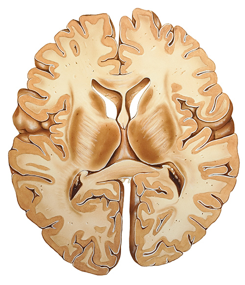
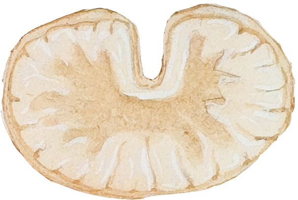
White matter refers to the areas of the CNS with the majority of axons, the long cords that extend from neurons. Most axons are coated in myelin – a white, fatty insulating cover that helps nerve signals travel quickly and reliably. In the brain, white matter is buried under the grey surface, carrying signals across different parts of the brain. In the spinal cord, white matter is the external layer surrounding the grey core.
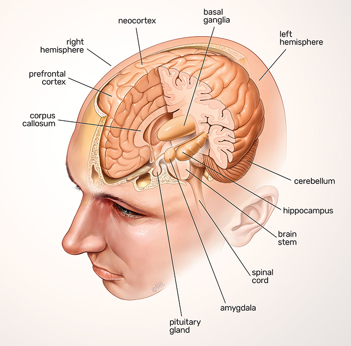
If the CNS is the processing center of the human body, the brain is its headquarters. It is broadly organized into three main regions: the forebrain, the midbrain, and the hindbrain. The largest of these three is the forebrain (derived from the prosencephalon in the developing brain). It contains the large outermost layer of the brain, the wrinkly cerebral cortex, and smaller structures towards its centre, such as the thalamus, hypothalamus, and pineal gland.
The midbrain (derived from the mesencephalon in the developing brain) serves as the vital connection point between the forebrain and the hindbrain. It’s the top part of the brainstem, which connects the brain to the spinal cord.
The hindbrain (derived from the rhombencephalon in the developing brain) is the lowest back portion of the brain, containing the rest of the brainstem made up of the medulla oblongata and the pons, and also the cerebellum – a small ball of dense brain tissue nestled right against the back of the brainstem.
NEURONS
Neurons (also called neurones or nerve cells) are the fundamental units of the brain and nervous system, the cells responsible for receiving sensory input from the external world, sending motor commands to our muscles, and transforming and relaying the electrical signals at every step in between. More than that, their interactions define who we are as people. Having said that, our roughly 100 billion neurons do interact closely with other cell types, broadly classified as glia (these may outnumber neurons, although it’s not known).
The creation of new neurons in the brain is called neurogenesis, and this can happen even in adults.
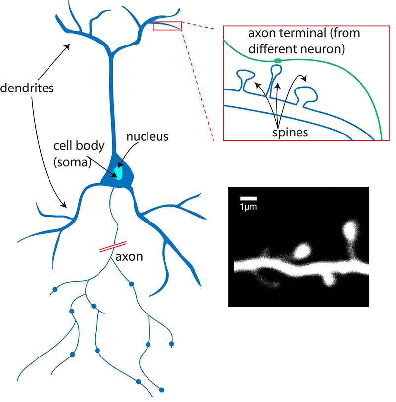
What does a neuron look like? A useful analogy is to think of a neuron as a tree. A neuron has three main parts: dendrites, an axon, and a cell body or soma (see image below), which can be represented as the branches, roots and trunk of a tree, respectively. A dendrite (tree branch) is where a neuron receives input from other cells. Dendrites branch as they move toward their tips, just like tree branches do, and they even have leaf-like structures on them called spines.
The axon (tree roots) is the output structure of the neuron; when a neuron wants to talk to another neuron, it sends an electrical message called an action potential throughout the entire axon. The soma (tree trunk) is where the nucleus lies, where the neuron’s DNA is housed, and where proteins are made to be transported throughout the axon and dendrites.
There are different types of neurons, both in the brain and the spinal cord. They are generally divided according to where they originate, where they project to and which neurotransmitters they use.
Axon – The long, thin structure in which action potentials are generated; the transmitting part of the neuron. After initiation, action potentials travel down axons to cause the release of neurotransmitters.
Dendrite – The receiving part of the neuron. Dendrites receive synaptic inputs from axons, with the sum total of dendritic inputs determining whether the neuron will fire an action potential.
Spine – The small protrusions found on dendrites that are, for many synapses, the postsynaptic contact site.
Action potential – A brief electrical event typically generated in the axon that signals the neuron as ‘active’. An action potential travels the length of the axon and causes the release of neurotransmitters into the synapse. The action potential and consequent transmitter release allow the neuron to communicate with other neurons.
PARTS OF THE BRAIN
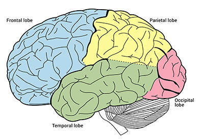
The brain’s cerebral cortex is the outermost layer that gives the brain its characteristic wrinkly appearance. The cerebral cortex is divided lengthwise into two cerebral hemispheres, each of which traditionally has been divided into four lobes: frontal, parietal, temporal, and occipital.
Although we now know that most brain functions rely on many different regions across the entire brain working in conjunction, it is still true that each lobe carries out the bulk of certain functions.
FOREBRAIN
By far the largest region of your brain is the forebrain (derived from the developmental prosencephalon), which contains the entire cerebrum and several structures directly nestled within it – the thalamus, hypothalamus, pineal gland, and the limbic system.
Cerebrum and the cerebral cortex. When you picture the iconic shape of the human brain, the majority of what’s visible is the cerebrum, with its wrinkly, pinkish-grey outer appearance. It makes up around 85% of the brain and consists primarily of grey matter, divided into two hemispheres.
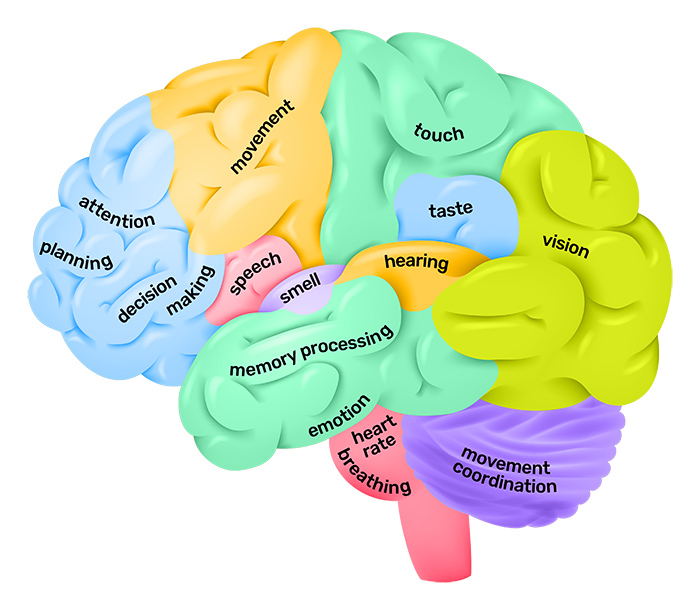
The cerebrum is where most of the important brain functions happen, such as thinking, planning, reasoning, language processing, and interpreting and processing inputs from our senses, such as vision, touch, hearing, taste, and smell.
The outer layer of the cerebrum is called the cerebral cortex, and in each hemisphere, it is traditionally divided into four lobes – frontal, parietal, occipital, and temporal. Communications between the two hemispheres are maintained by a fibrous bridge called the corpus callosum, which is formed in utero.
Beneath the surface of the hemispheres are large knots of neurons called the basal ganglia, which specialize in programming and executing our motor functions. When the basal ganglia are affected by diseases such as Parkinson’s, patients have tremors and uncontrolled movements.
Control centres for making sense of our bodies. Apart from the cerebrum, the forebrain also contains several small but highly important structures located towards the centre of the brain and are included in the limbic system. Collectively, these are called the diencephalon, and they regulate things like the body’s sensory perception, motor functions, and hormones.
The thalamus consists of two lobes of grey matter tucked away right under the cerebral cortex. It is a prime processing centre for sensory information, as it links up the relevant parts of the cerebral cortex with the spinal cord and other areas of the brain important for our senses. The thalamus also controls sleep.
The hypothalamus is quite small, only about the size of an almond. As its name suggests, it can be found right underneath the thalamus, and despite its small size, it is the major control centre of the autonomic motor system. It is involved in some hormonal activity and connects the hormonal and nervous systems. The hypothalamus also works to regulate blood pressure, body temperature, and overall homeostasis.
The pineal gland is even smaller than the hypothalamus – only about the length of a grain of rice – and is tucked between the two lobes of the thalamus. It is shaped like a tiny pine cone, and its main job is to produce the hormone melatonin, which regulates our sleep-wake cycles. Just like the hypothalamus, it is also involved in regulating hormonal functions.
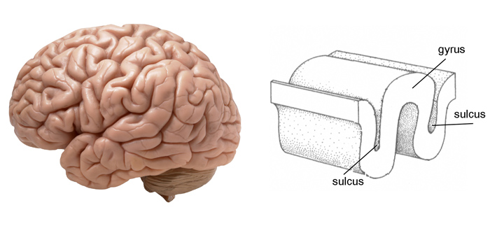
Bumps and grooves of the brain. In humans, the lobes of the brain are divided by bumps and grooves. These are gyri (bumps) and sulci (grooves or fissures). The folding of the brain and the resulting gyri and sulci increase its surface area and enable more cerebral cortex matter to fit inside the skull.
Frontal Lobe. The frontal lobe is separated from the parietal lobe by a space called the central sulcus and from the temporal lobe by the lateral sulcus.
The frontal lobe is generally where higher executive functions, including emotional regulation, planning, reasoning, and problem-solving, occur. This is why in frontotemporal dementia, personality changes are often the first signs of the disease.
The most famous case of frontal lobe dysfunction is the story of railway worker Phineas Gage. In 1848, Gage was using a tamping iron to pack in gunpowder for blasting a tunnel through rock. While his head was slightly turned, a mistaken strike sparked an explosion that forced the rod upwards into his left eye and out through his skull.
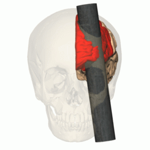
Miraculously, Gage survived, blinded in his left eye and sustaining damage to much of his left frontal lobe. After the accident, others noticed changes in Gage’s personality: before the accident, he was known as responsible and hard-working, but afterward, he became disrespectful and foul-mouthed and had difficulty carrying out plans.
The frontal lobe also contains the primary motor cortex, the major region responsible for voluntary movement.
Parietal lobe. The parietal lobe is behind the frontal lobe, separated by the central sulcus. Areas in the parietal lobe are responsible for integrating sensory information, including touch, temperature, pressure, and pain.
Because of the processing that occurs in the parietal lobe, we can, for example, discern from touch alone that two objects touching the skin at nearby points are distinct rather than one object. This process is called two-point discrimination. Different areas of the body have more sensory receptors and so are more sensitive than others in discerning distinct points. Using callipers or a folded paperclip and asking a subject to keep their eyes closed, this test can be used to check parietal lobe function.
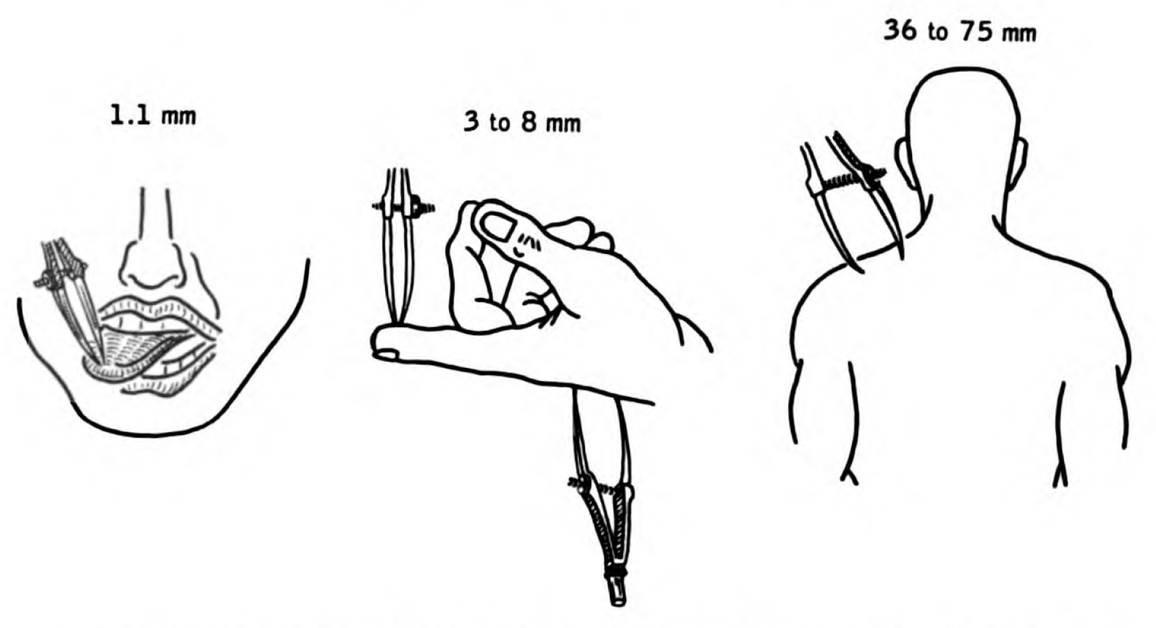
While a subject’s eyes are closed, a folded paper clip can be used to test two-point discrimination, which is mediated by the parietal lobe. The tester alternates using one point and two points on the area being tested (e.g. finger, shoulder, arm). The subject is asked to report whether they felt one or two points.
Temporal lobe. Separated from the frontal lobe by the lateral fissure, the temporal lobe also contains regions dedicated to processing sensory information, which is particularly important for hearing, recognizing language, and forming memories.
Auditory Processing. The temporal lobe contains the primary auditory cortex, which receives auditory information from the ears and secondary areas and processes the information so we understand what we’re hearing (e.g. words, laughing, a baby crying).
Visual Processing. Certain areas in the temporal lobe make sense of complex visual information, including faces and scenes.
Memory. The medial (closer to the middle of the brain) temporal lobe contains the hippocampus, a region of the brain important for memory, learning, and emotions.
Occipital lobe. The occipital lobe is the major visual processing centre in the brain. The primary visual cortex, also known as V1, receives visual information from the eyes. This information is relayed to several secondary visual processing areas, which interpret depth, distance, location, and the identity of seen objects.
LIMBIC SYSTEM. The limbic system is the part of the brain involved in our behavioural and emotional responses, especially when it comes to behaviours we need for survival: feeding, reproduction, caring for our young, and fight or flight responses.
You can find the structures of the limbic system buried deep within the brain, underneath the cerebral cortex, and above the brainstem. The thalamus, hypothalamus (production of important hormones and regulation of thirst, hunger, mood, etc.), and basal ganglia (reward processing, habit formation, movement, and learning) are also involved in the actions of the limbic system, but two of the major structures are the hippocampus and the amygdala.
Hippocampus. The hippocampus, like many other structures in the brain, comes as a pair, one in each hemisphere of the brain. It resembles the shape of a curvy seahorse (and is named after its scientific genus), and is essentially the memory centre of our brains. Here, our episodic memories are formed and cataloged to be filed away in long-term storage across other parts of the cerebral cortex.
Connections made in the hippocampus also help us associate memories with various senses (the association between Christmas and the scent of gingerbread would be forged here). The hippocampus is also important for spatial orientation and our ability to navigate the world.
The hippocampus is one site in the brain where new neurons are made from adult stem cells. This process is called neurogenesis and is the basis of one type of brain plasticity. So it’s not surprising that this is a key brain structure for learning new things.
Amygdala. The amygdala’s name refers to its almond-like shape. Located right next to the hippocampus, the left and right amygdala play a central role in our emotional responses, including feelings like pleasure, fear, anxiety, and anger. The amygdala also attaches emotional content to our memories, and so plays an important role in determining how robustly those memories are stored. Memories that have strong emotional meanings tend to stick.
The amygdala doesn’t just modify the strength and emotional content of memories; it also plays a key role in forming new memories specifically related to fear. Fearful memories are formed after only a few repetitions. This makes ‘fear learning’ a popular way to investigate the mechanisms of memory formation, consolidation, and recall.
Suppressing or stimulating activity in the amygdala can influence the body’s automatic fear response, which kicks in when something unpleasant happens, such as a startling noise. Receptors have been discovered in the amygdala that could help to develop new types of anti-anxiety drugs.
New neurons are also made in the amygdala.
HINDBRAIN
The hindbrain (developmentally derived from the rhombencephalon) is one of the three major regions of our brains, located at the lower back part of the brain. It includes most of the brainstem and a dense, coral-shaped structure called the cerebellum. The brainstem is one of the most important parts of the entire central nervous system because it connects the brain to the spinal cord and coordinates many vital functions, such as breathing and heartbeat.
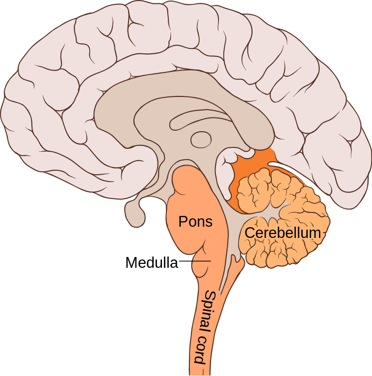
There are three main parts of the hindbrain: the pons, the cerebellum, and the medulla oblongata. Most of the 12 cranial nerves are found in the hindbrain.
Pons. The pons gets its name from the Latin word for ‘bridge”, and it connects the rest of the brainstem to the cerebral cortex. Bulbous in shape, it sits right underneath the midbrain and serves as a coordination centre for signals and communications that flow between the two brain hemispheres and the spinal cord.
Four cranial nerves are found in the pons: the abducens nerve helps coordinate eye movement; the facial nerve coordinates movement and sensation in the face; the vestibulocochlear nerve processes sound and helps us maintain balance; and the trigeminal nerve coordinates chewing and carries sensory information from the face and the head.
Cerebellum. Behind the pons and the rest of the brainstem sits a structure called the cerebellum (Latin for ‘little brain’). In cross-section, this part looks like a layered, wrinkly coral. Just like the cortex, it has two hemispheres, with a dense layer of grey matter surrounding an inner region of white matter. It also contains special neurons called Purkinje cells, which process many signals at once due to their highly complex dendrite branches.
The cerebellum coordinates our sensations with responses from our muscles, enabling most of our voluntary movements. It also processes nerve impulses from the inner ear and coordinates them with muscle movement, thus helping us maintain balance and posture.
Medulla oblongata. The lower part of both the brainstem and the overall hindbrain is the medulla oblongata, where the brain transitions to the spinal cord. It is only about 3cm long, but the medulla is an indispensable nerve tract that contains the control centres for our autonomic vital functions – heart rate, blood pressure, breathing – and many involuntary reflexes such as swallowing and sneezing.
The medulla contains both white and grey matter, and four cranial nerves stem from this region: the glossopharyngeal nerve coordinates some taste sensations and mouth movements; the vagus nerve controls mouth movements, voice, and the gag reflex; the accessory nerve coordinates head and neck movements; and the hypoglossal nerve controls tongue movements and muscles involved in our speech.
AN ATLAS OF THE BRAIN
Cataloguing its components may help understand how it works

Lord Rutherford, the discoverer of the atomic nucleus, divided science into physics and stamp collecting. (He was, after all, a physicist.) But he had a point. Other sciences, such as astronomy, chemistry, geology and, most notably, biology, rely a lot on collecting things (not literally, in the case of astronomy) and classifying them in various ways that would delight philatelists. Physics, by contrast, relies on analyzing phenomena.
That said, the philatelist branches of science have been pretty successful, especially biology. And this week sees the addition of a new album to biology’s collection, in the form of 21 papers about the brain and its cells. The work was done under the purview of the Brain Initiative Cell Census Network, which is organized by the National Institutes of Health in America. The papers are published in various bits of the American Association for the Advancement of Science’s empire of journals, Science and its spin-offs. They are intended to help answer three, related questions: what and where are a brain’s cellular components; which cells are involved in neurological and psychiatric illnesses; and what makes the brains of Homo sapiens different from those of other animals?
Brains, particularly human ones, are the most complex objects in the known universe. That complexity is emphasized by the fact that, as these papers confirm, they are reckoned (depending on how you define such things) to contain about 3,000 different types of cells. For comparison, it was not so long ago that entire human bodies, brains included, were estimated to be built from just 300 cell types.
The estimate was confirmed by a team of researchers at the Karolinska Institute in Stockholm and the Allen Institute for Brain Science in Seattle. They studied, post-mortem, the brains of three men and a woman, taking 606 samples from nine regions of the organ and also one from the spinal cord. They then extracted the nuclei of individual cells and looked at the RNA molecules therein.
RNA is chemically similar to DNA and comes in many varieties, each with a different job. One of the most important is to act as a messenger, carrying instructions for how to make particular proteins from the nucleus, where the genes that encode protein recipes are stored as DNA, to the cellular factories, called ribosomes, which churn those proteins out. Analyzing those RNA messengers reveals which proteins are being made. And the proteins a cell produces determine what type of cell it is.
On that basis, the two institutes’ researchers found 31 “superclusters” of cells with similar patterns of RNA expression. These, in turn, are divided into 461 clusters and 3,313 subclusters—in effect, individual cell types. Superclusters tended to be concentrated in one or a few brain regions (for example, the cerebral cortex had 16, the hippocampus had 12, and the cerebellum had six). But there was an exception. This was a supercluster that the team called “splatter neurons”. These turned up all over the place.
Other groups dug into the details. Bing Ren of the University of California, San Diego and his colleagues, for example, attempted to characterize cells not directly by their messenger RNA but rather by differences in the way the DNA in their chromosomes was packaged. By allowing—or forbidding—access to the DNA, such packaging helps regulate which genes are transcribed into RNA messengers.
This yielded 107 recognizable patterns. Intriguingly, some of these patterns could be correlated with neurological traits and illnesses. The team compared the locations of pieces of DNA that control gene transcription and which are known to have disease-associated variants with the DNA-packaging patterns of different sorts of cells. They found correlations with 19 conditions, including schizophrenia, depression, bipolar disorder, Alzheimer’s disease, and various forms of addiction.
A third group, meanwhile, led by Rebecca Hodge and Trygve Bakken of the Allen Institute, compared the brains of humans with those of chimpanzees, gorillas, macaques (a group of old-world monkeys) and marmosets (a group of new-world monkeys), looking for clues to the elusive question of what makes human brains human. They found that part of the answer may lie not in the neurons themselves but in the supporting cast of non-neuronal brain cells called glial cells.
Clues to humanity’s essence are likely to be found in parts of the genome called hars and hcondels. hars stands for “human accelerated regions”. These are bits of the genome that have remained unchanged in apes and monkeys but are altered in humans. hcondels—“human-conserved deletions”—are the opposite: places where DNA found in apes and monkeys is missing in people. The team found that genes in and around both areas are often particularly active or inactive in glial cells.
Glial cells come in three varieties: astrocytes, which regulate the flow of information across junctions between neurons; microglia, which prune links between neurons to keep the network in order; and oligodendrocytes, which insulate nerve fibres and tweak signals running along them. That suggests part of what gives human brains their humanness lies in these microscopic details of their architecture.
A good start, then. But this work only scratches the surface of the brain’s true complexity. Understanding that properly requires not only listing and describing the various components but also elucidating how they are wired together into functional units. Collecting stamps is fine. But you need more than that to work out how a postal service operates.
